


BY: AMANDA FORESTIERI
Individuals who suffer from mild to moderate brain injuries often have long-lasting debilitating symptoms, despite the brain appearing ‘normal’ on structural MRI or CT scans.
BIST Social Work placement student Amanda Forestieri, sat with neuroscientist Dr. David Corey, to discuss his work with functional Magnetic Resonance Imaging (fMRI).
Dr. Corey has worked with and treated individuals with chronic pain, PTSD, and mild to moderate TBIs for 40 years. He has worked in interdisciplinary teams, and with many patients who have struggled to prove they are experiencing TBI symptoms. Dr. Corey says this is likely due to metabolic changes in the brain, or changes which are too microscopic for a structural MRI or CT scan to pick up.
Functional MRI’s versus MRI
The functional MRI (fMRI) focuses on oxygen atoms, to give a rough measure of the metabolic activity of the cells in the brain. The fMRI looks primarily at how the brain is functioning, whereas the standard MRI assesses structure only (eg. tumors).
How the FMRI Works
The fMRI detects the blood flow in a particular area of the brain when the patient is asked to perform a task. Doctors are able to see if it functions in the same way as in a brain without an injury. Individuals with a brain injury are tested in an activation paradigm, meaning that the individual is asked to perform a task in the scanner as opposed to a resting state. The task is called the Tower Task, and individuals are asked to sort coloured balls in different containers on a screen.
Dr. Corey has noticed different activation in the brain of someone with a brain injury. The brain injury population loses synchronicity between the two hemispheres, and there is more activation in certain parts of the brain compared to a non-injured brain when performing a task. A statistical test is then done to determine whether the patient’s brain function falls into the “normal” or “abnormal” range. This image displays a control groups’ scan vs. someone with PCS (Post Concussion Syndrome).
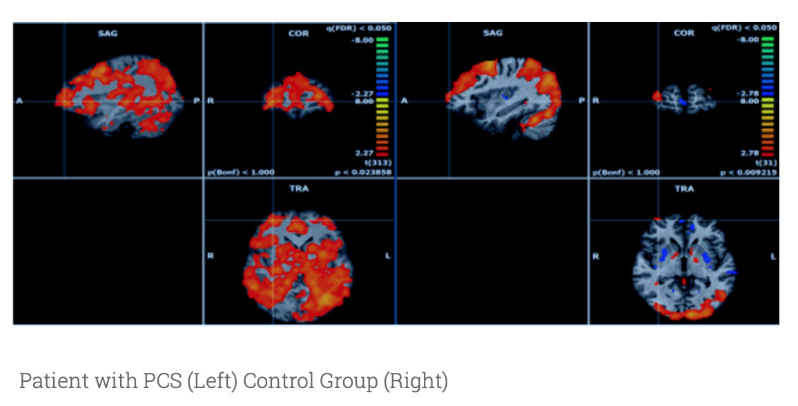
The above shows widespread activation in the brain of a patient with PCS. More research is being done as to why this is the case.
The fMRI can be used as a tool to show evidence of brain injury and clarify diagnosis. It is another tool to show verification that brain function is “abnormal” in a person who experienced a mild to moderate brain injury.
In order for individuals to prepare for an fMRI, one needs to be medically cleared for a standard MRI and be able to lie still for at least 10 minutes at a time. Claustrophobia is something else to keep in mind, as the fMRI is in a closed environment. To conduct an fMRI, patients can’t take medication, such as tranquilizers, before the scan, but many people can learn some calming techniques to manage anxiety.
What is fMRI currently being used for?
FMRI equipment is expensive, and its analysis is highly complex. Currently, few doctors are trained to understand the data. It is not funded by OHIP at present, nor is it used widely for clinical purposes.
Having said that, Dr. Corey believes that in the future the fMRI may be brought forward as a clinical tool.
“When you produce an objective measure of something, people pay attention to it, especially in the medical profession,” Dr. Corey said. As of right now, fMRIs are mostly being used in a medical-legal context. However, it is exciting to think about the technology that one might see in the future for those with a mild to severe brain injury, allowing those, individuals to receive better diagnosis and treatment. More information is available on Dr. Corey’s work and his contact information on www.brainscaninc.com.
Amanda Forestieri is a passionate 4th-year social work student who hopes to work in social services. When Amanda is not in school, you can find her reading a good book, going for walks or singing and creating music with others. She wants to one day be known as the singing social worker!